The Core Idea of the Vision Banana Paper

The project notes summarize Image Generators are Generalist Vision Learners around one central question: whether strong image generators already contain rich visual structure priors that can be released through lightweight instruction tuning.

Idea 01

Image generation pretraining as vision pretraining

The paper assumes that high-quality generators have learned boundaries, spatial relations, scale, material, geometry, and semantics. Instruction tuning may expose those internal priors as vision-understanding ability.

Idea 02

Parameterize outputs as RGB structure maps

Semantic segmentation, instance segmentation, depth, surface normals, and referring segmentation are unified as generated, decodable result images: input image plus prompt, generated map, then postprocessed structure.

Idea 03

Control outputs with language and schema

Vision Banana can specify targets and colors through natural language, JSON, RGB tuples, or hex colors. In medicine, this must become controlled terminology, fixed palettes, and reproducible protocols.

How It Differs from Other Image Segmentation Models

In plain terms, conventional segmentation models behave like pixel-level classifiers: each pixel is judged as target, background, or a class label. Vision Banana-style methods ask an image generator to draw a decodable answer map: target regions, instances, depth, or normals are rendered with specified colors and decoded afterward.

Typical segmentation-model implementation Image -> feature encoder -> segmentation head -> probability map -> thresholded mask The core is discriminative prediction: pixels are classified, usually with direct supervision on logits or masks.
Vision Banana-inspired implementation Image + prompt + color schema -> generated RGB structure map -> decoded mask / depth / normal The core is generative prediction: the model outputs a new answer image, turning vision tasks into image generation.
Dimension
Traditional / modern segmentation models
Vision Banana-style generative models
Model interface
An image is passed to a segmentation head or mask decoder that outputs logits or masks.
An image and prompt condition a generator that produces RGB masks, depth maps, normal maps, or other structure maps.
Output form
Usually class probabilities, binary masks, multiclass masks, or instance masks as tensor-like outputs.
A human-readable generated image is decoded through a color codec, thresholds, and connected components.
Task boundary
Most models optimize segmentation; SAM, MedSAM, SAT, VISTA3D, and related models extend promptable segmentation.
The same generation interface attempts to cover segmentation, geometry, depth, surface normals, measurements, and uncertainty maps.
Medical risk
Strengths include coordinate registration, efficiency, and quantifiable supervision, but open vocabulary and absent targets still need design.
Strengths include a unified interface and generative priors; risks include color drift, global offset, local deformation, and hallucinated anatomy.
Research value
These are strong baselines for supervised medical image-mask segmentation.
The key question is whether generative pretraining can become a reliable medical dense-prediction paradigm, and where it fails.
A Feasibility Study of Vision Banana-style Methods for Medical Imaging

This page summarizes several GPT-Images-2 probes. The natural portrait case produced a detailed RGB mask, but medical images showed registration drift and anatomical mismatch that are unacceptable for serious medical segmentation.

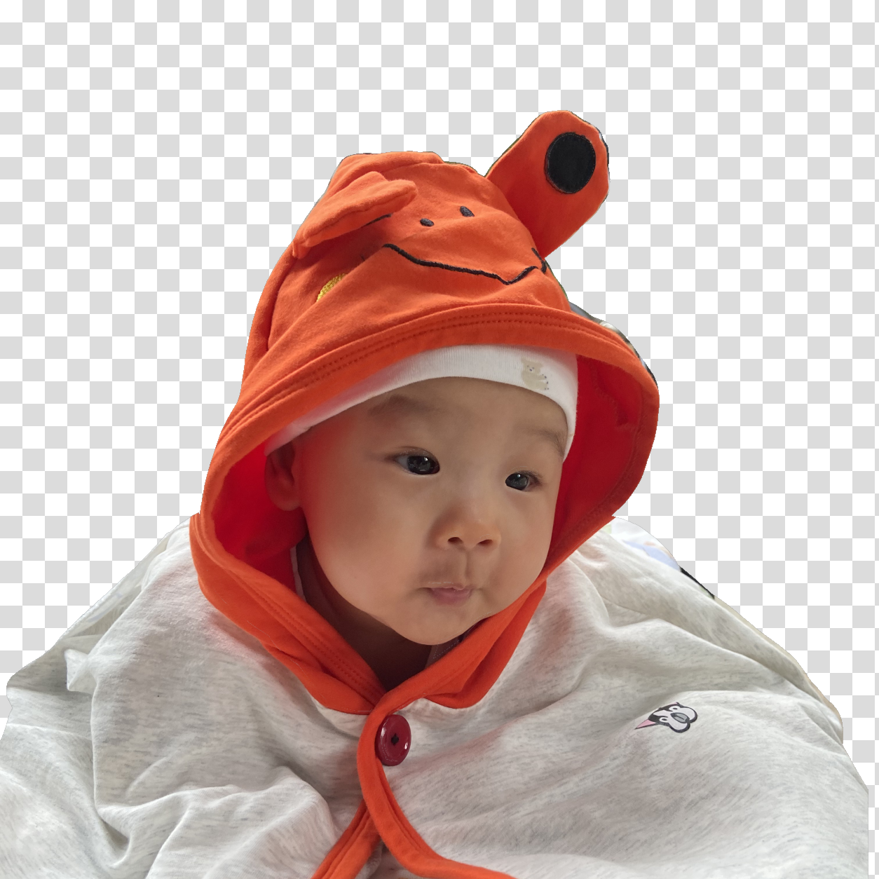
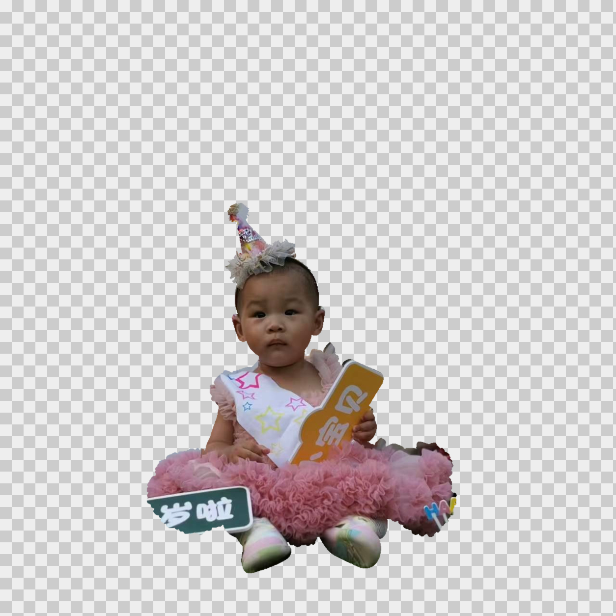
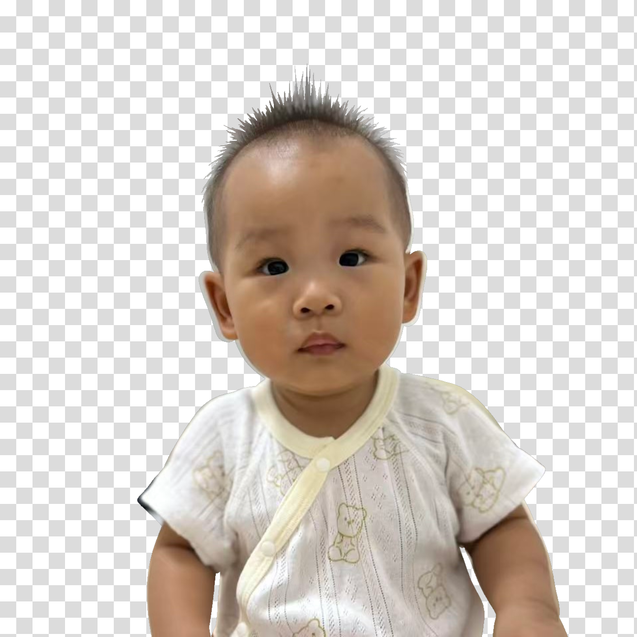
Aligned cutout preview for the natural portrait
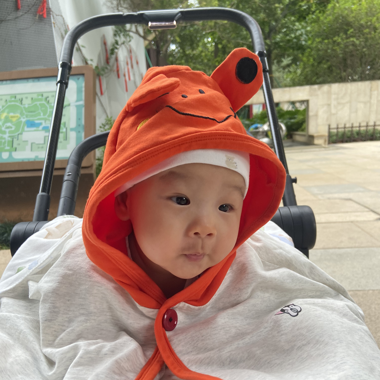
Natural image: fine contours, registration needed
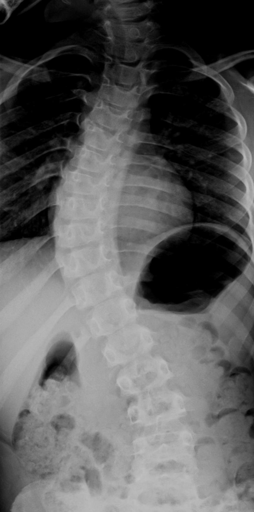
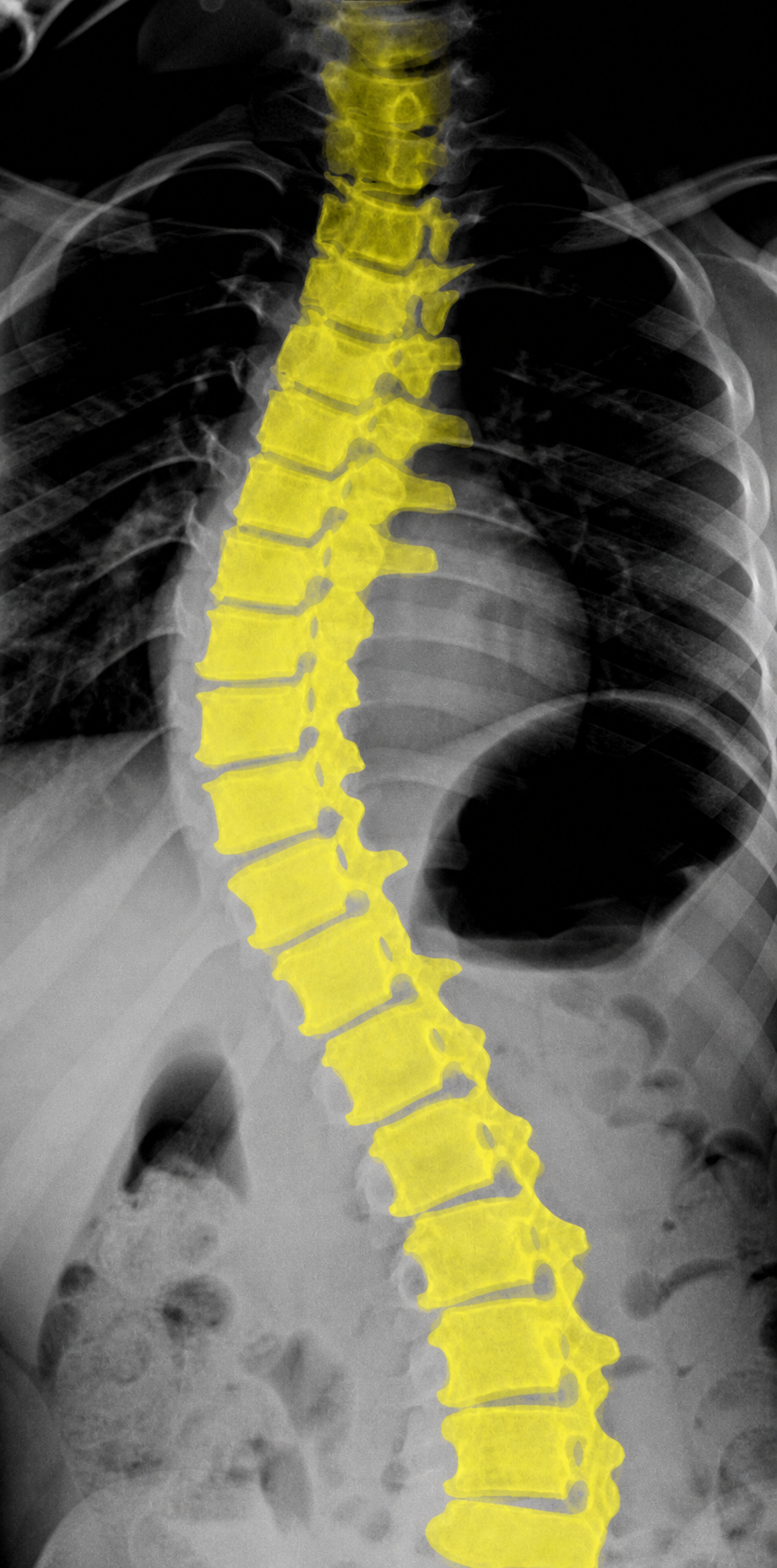
Failed GPT overlay for the spine X-ray
X-ray: generated structure is not source-registered
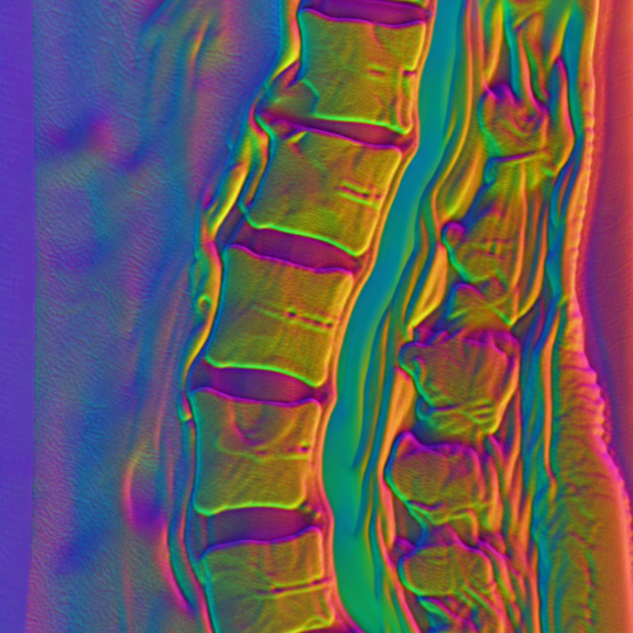
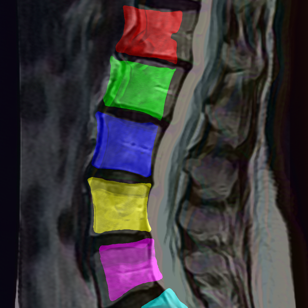
Surface normal-style map for the spine MRI
MRI: normal-style visualization has research value
Natural Image Usable after registration Simple cases dx -15 to -24px; complex case approx. dx -5px, dy +25px
X-ray Failed Generative hallucination and coordinate drift
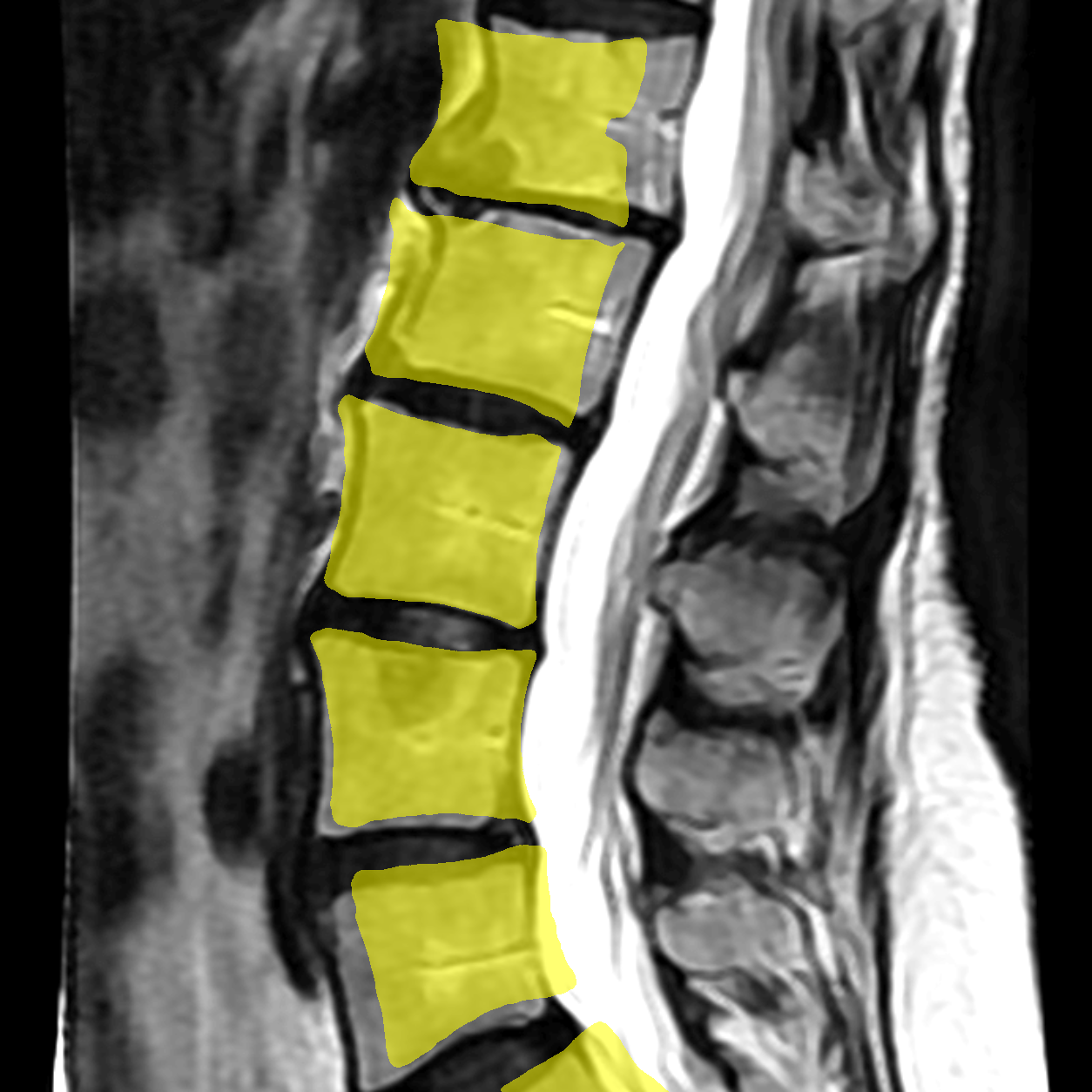
MRI Segmentation Below medical requirements Vertebral boundaries differ from the source
Surface Normal Useful visualization Promising as a structural representation probe

How the Paper's Method Was Transferred to This Project

The key Vision Banana idea is to reframe vision tasks as image generation rather than traditional logit prediction: input an image and prompt, then output a visual, decodable RGB structure map.

01

Image generators as vision learners

Use boundaries, geometry, spatial relations, and semantic structure learned during image generation pretraining, then expose dense prediction through instruction tuning.

02

Vision outputs parameterized as RGB

Semantic segmentation, instance segmentation, depth, and surface normals can all be represented as RGB result images and decoded into structured outputs.

03

Prompt plus palette codec

Text specifies the target and color schema, such as background = black and target = yellow, followed by color projection and connected-component analysis.

04

Reliability constraints for medicine

Medical tasks must additionally handle absent targets, 3D continuity, uncertainty, color drift, and hallucinated anatomy. Visual appeal is not enough.

Medical Image
Prompt / RGB Schema
GPT-Images-2 Structure Map
Decode and Evaluate

Test Images and Results

The images below come from the project directory. Failed examples are intentionally kept because they decide whether this direction is ready for serious medical research.

Case A

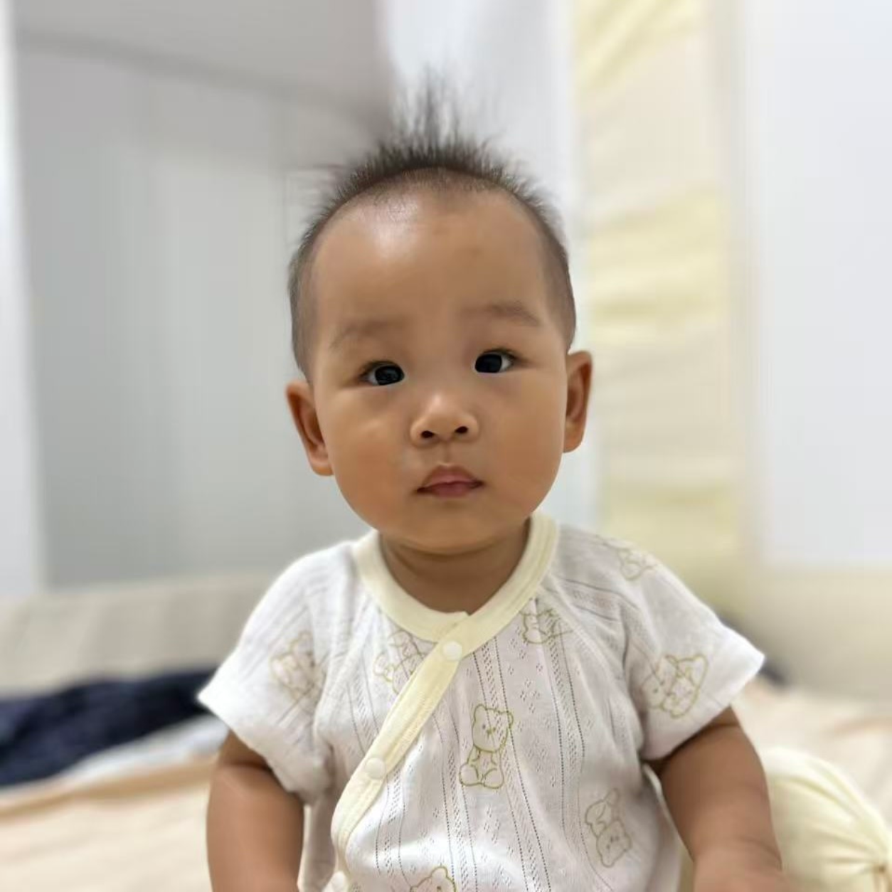
Natural portrait baby_1254.png: clean person extraction on a simple background

Original baby_1254 source image
Source image
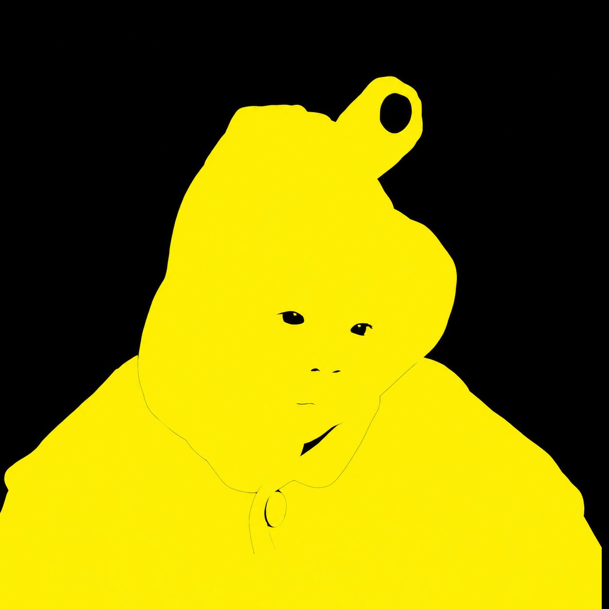
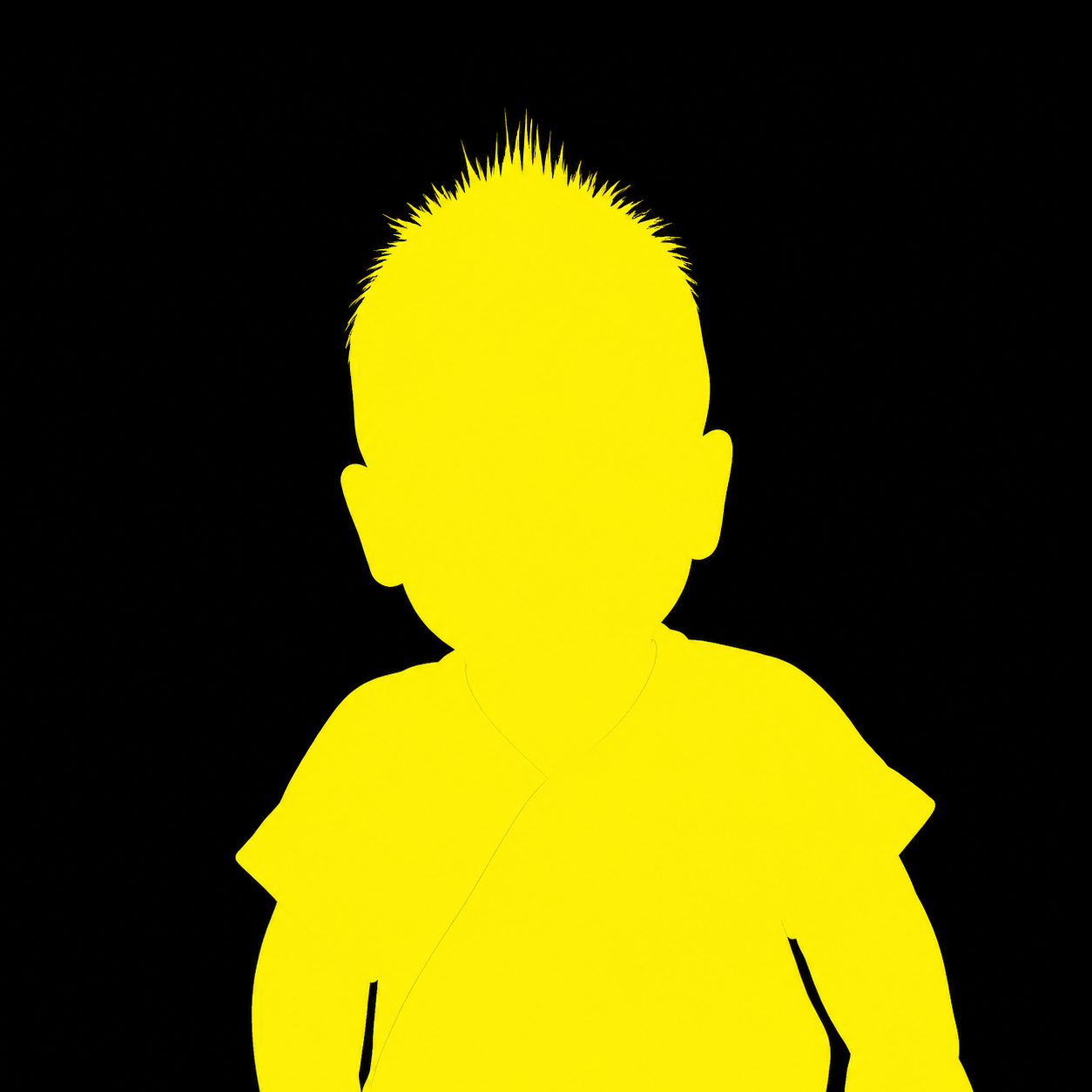
Aligned RGB mask for baby_1254
RGB mask, aligned
Transparent-background cutout preview for baby_1254
Transparency check

This case acts as a positive control for the natural-person task: both the source image and GPT output are 1254 x 1254. The mask contour is usable, the estimated offset is dx=-15px, dy=+50px, and the aligned result produces a clean transparent PNG.

Case B

Natural portrait baby2_1254.png: strong contours, global offset

Original indoor baby portrait
Source image
Aligned RGB mask for the baby portrait
RGB mask, aligned
Transparent-background cutout preview for baby2_1254
Transparency check

The GPT-Images-2 mask responds well to the person boundary and fine hair details. The main issue is not contour quality, but global displacement. The estimated offset is dx=-24px, dy=+52px.

Case C

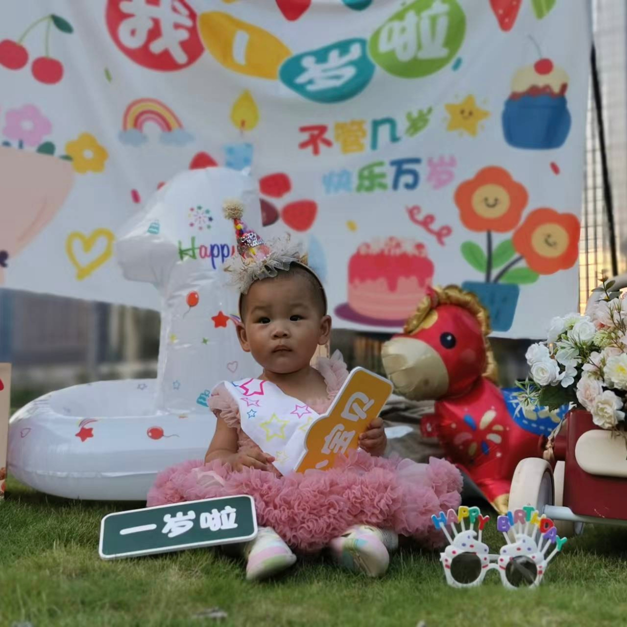
Complex birthday scene baby2_1254.jpg: occluding props must remain, offset changes

Original complex birthday-scene JPG
Source image
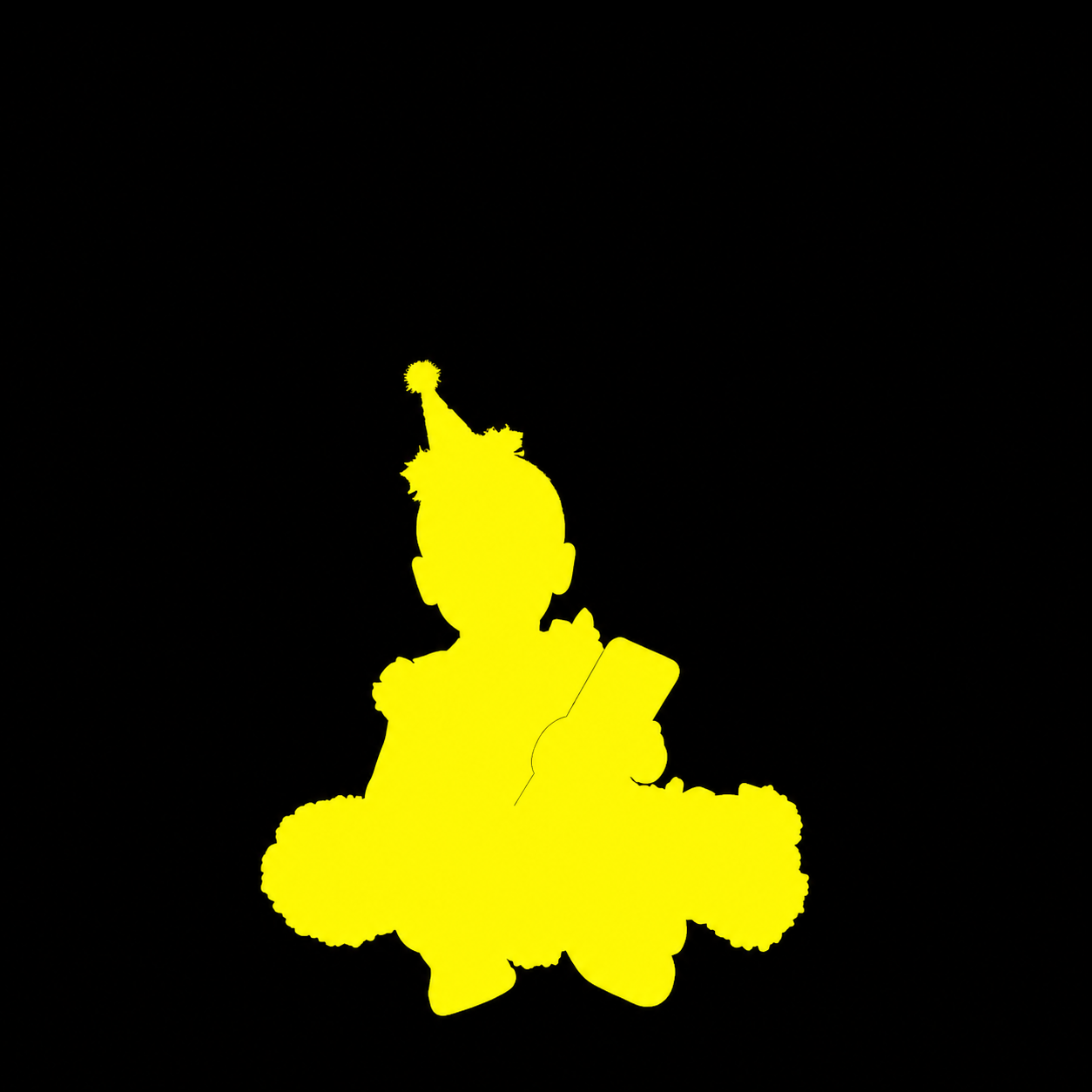
Aligned RGB mask for the complex JPG case
RGB mask, aligned
Transparent-background cutout preview for the complex JPG case
Transparency check

This image is harder than the first two natural-person cases: the hand sign and foreground sign are real occluders in front of the child, so removing all props would contradict the visible image. The selected result uses the second-best mask that preserves foreground occlusion, with a re-estimated offset of dx=-5px, dy=+25px. It is useful for natural-image probing, but already requires judgment and postprocessing.

Case D

Spine X-ray: not source-coordinate registered

Original spine X-ray
Source image
GPT-generated spine X-ray overlay
GPT overlay

The result resembles a medical structure, but it is not a strict segmentation in the source-image coordinate system. It behaves more like a visually plausible spine shape, which is unacceptable for medical segmentation.

Case E

Spine MRI RGB mask: vertebrae visible, boundaries unusable

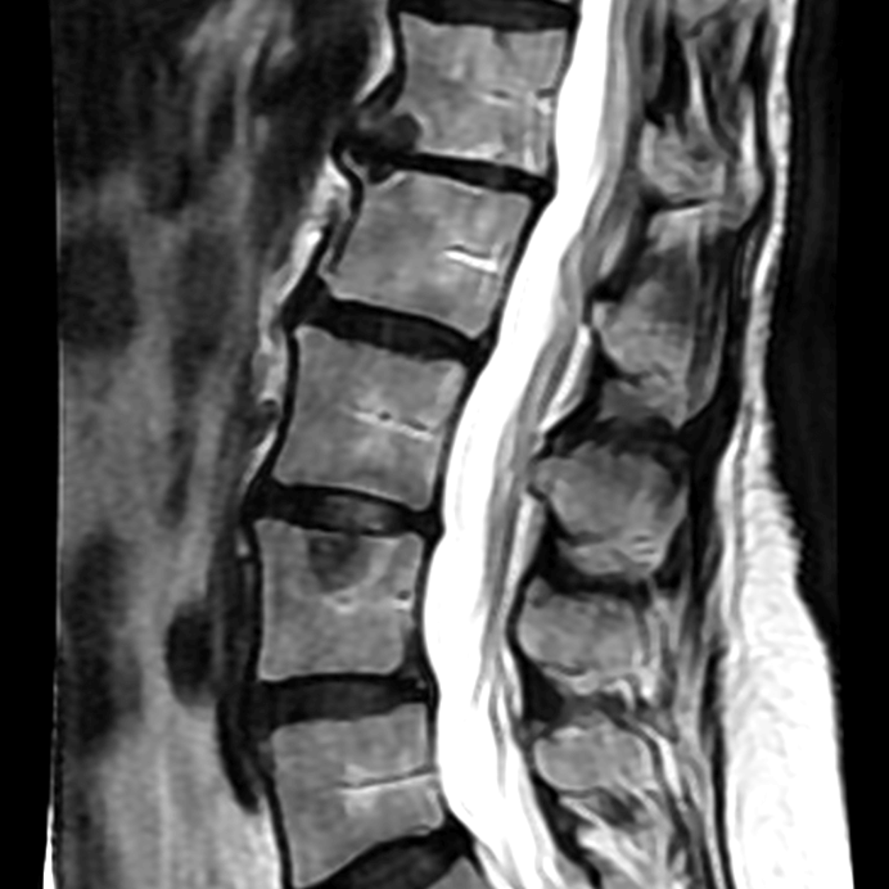
Original spine MRI
MRI source image
Spine MRI RGB mask overlay
GPT mask overlay

Compared with the X-ray case, vertebral block structures in the MRI are easier for the model to capture. However, after overlaying the result, vertebral edges, lower anatomy, and source-image boundaries still differ too much to serve as a medical mask.

Case F

Surface normal-style map: structural enhancement, not ground truth

GPT surface normal-style map
Normal-style map
Palette-constrained normal-style postprocessed image
Palette plus normal-style

The normal-style output enhances structures around vertebrae, discs, and the spinal canal, but it is not a physical surface normal. The palette-constrained version combines a GPT mask with a GPT normal map through postprocessing; it is not a single pure GPT output.

Why the Medical Conclusion Is Conservative

Good-looking natural-image contours do not imply medical usability. Medical imaging requires verifiable coordinates, boundaries, structure existence, and uncertainty.

Errors that medicine cannot accept

  • Global structural displacement that cannot be fixed by visual inspection alone.
  • Plausible but nonexistent anatomical structures.
  • Vertebral boundaries that are smoothed, summarized, or redrawn.
  • Output colors that are not a strict palette and require projection plus threshold decoding.

Interim judgment from this project

GPT-Images-2 is useful for rapidly probing the Vision Banana paradigm, but zero-shot medical-image outputs should not be used as main paper results. Deeper research should convert the idea into a reproducible training framework and quantitative evaluation protocol.

Qualitative Rating

Natural portrait contourGood
X-ray registrationFailed
MRI vertebra recognitionPartial
Clinical usabilityInsufficient

Current Limitations: Why This Is Not Yet a Full Paper

The current results are suitable as a preliminary feasibility study and failure-mode note, but they are not enough to support a full medical-imaging paper. The following evidence is still missing.

Insufficient sample size

The current work is mainly case-based. A paper-level claim needs tens to hundreds of medical images across modalities, acquisition quality, and anatomical variation.

No expert ground truth masks

Medical segmentation must be compared against expert annotations. Without ground truth, registration drift, boundary error, and missing structures cannot be measured rigorously.

No quantitative metrics

The study needs Dice, IoU, Hausdorff distance, boundary error, centroid displacement, color decoding error, and failure rate, rather than visual inspection alone.

Protocol not fixed yet

Prompts, output size, RGB palette, threshold decoding, connected-component filtering, and whether post-hoc translation is allowed must be fixed before evaluation.

No baseline comparison

The method should be compared with conventional medical segmentation models, general-purpose segmentation models, and generative structure-map baselines.

Failure modes need statistics

Observed failures include global offset, local deformation, occlusion handling, generative completion, and anatomical hallucination; their frequency and causes need dataset-level analysis.

Next Research Directions

If this becomes a paper project, the current results should not be packaged as a success demo. The value is in designing rigorous methods around the observed failure modes.

Medical-domain instruction tuning

Train generative structure-map outputs on real medical image-mask datasets instead of relying directly on zero-shot general-model behavior.

Decodable RGB codec

Add palette regularization, color projection, connected components, and uncertainty-zone labels so color drift does not corrupt masks.

3D consistency

Extend from 2D slices to 2.5D, tri-plane, or 3D volumes, constraining structural continuity across adjacent slices.

Absent targets and uncertainty

Explicitly train empty masks, p_exist, and sampled uncertainty to reduce the most dangerous false-positive structures in medical scenarios.

References

Vision Banana project page: vision-banana.github.io · Paper: Image Generators are Generalist Vision Learners